Vision Center - Clarity Starts Here
Guidance and resources for vision health, eye care, and personalized eyewear. Matched with top eye doctors for optimal sight. Cutting through confusion to clarify your vision path.
Insights for Better Vision
Simplifying Every Aspect of Eye Care


We shine a spotlight on critical eye health subjects - translating complex topics into accessible guidance. Our goal is to explore vision care from all angles, providing clarity to support better eye and sight outcomes.

Vision Center - See and Be Seen
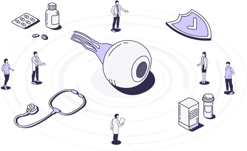
At Vision Center, we know that eyes are the window to the world - they shape how you experience life's beauty and connect with loved ones. Clear vision fosters wonder and brings joy into focus, making moments more colorful, vivid, and memorable. Good sight literally brightens life's views, while vision challenges can dim the details.
That’s why we exist - to guide and empower people to truly see and be seen.
Our team of eye doctors, optometrists, and patient advocates deeply understand the challenges of navigating vision care. We research, create, and curate practical vision health resources you can trust.
Vision Center offers reliable guidance to answer your questions around eye conditions, finding eyewear, connecting with eye doctors, and considering vision correction options if needed. We aim to bring life’s most meaningful moments into sharper focus.
All About LASIK
Latest Articles
Black Friday & Cyber Monday Glasses & Contacts Deals
Save on glasses and contacts this Black Friday and Cyber Monday. Compare the best deals from top eyewear retailers and maximize your holiday...
March 21, 2026
4 min read
Holiday Gift Guide for Glasses and Contacts Wearers
Find the best holiday gifts for glasses and contacts wearers. Practical gift ideas and top retailers to make your shopping easy and personal...
March 21, 2026
3 min read
What to Know About Astigmatism After LASIK
LASIK can treat astigmatism, but some people still notice residual or returning astigmatism after surgery.
March 21, 2026
7 min read
Best Optometry Schools in the U.S.
These are some of the best optometry schools in the U.S. to compare if you care about board outcomes, clinical training, externships, and re...
March 21, 2026
3 min read
When Would You Need Glasses After LASIK?
Many people see well without glasses after LASIK, but some still need glasses later because of healing changes, residual prescription, or pr...
Vision Center Podcast
Join us as we dive deep into the world of eye care, share inspiring stories, and explore the latest advancements in vision science. This Vision Center podcast is where vision health comes into focus. Our goal is to empower through education so you can access the vision care needed to see your best life.