Updated on February 20, 2024
Orbital Bone Functions and Fractures


Vision Center is funded by our readers. We may earn commissions if you purchase something via one of our links.
What is the Orbital Bone?
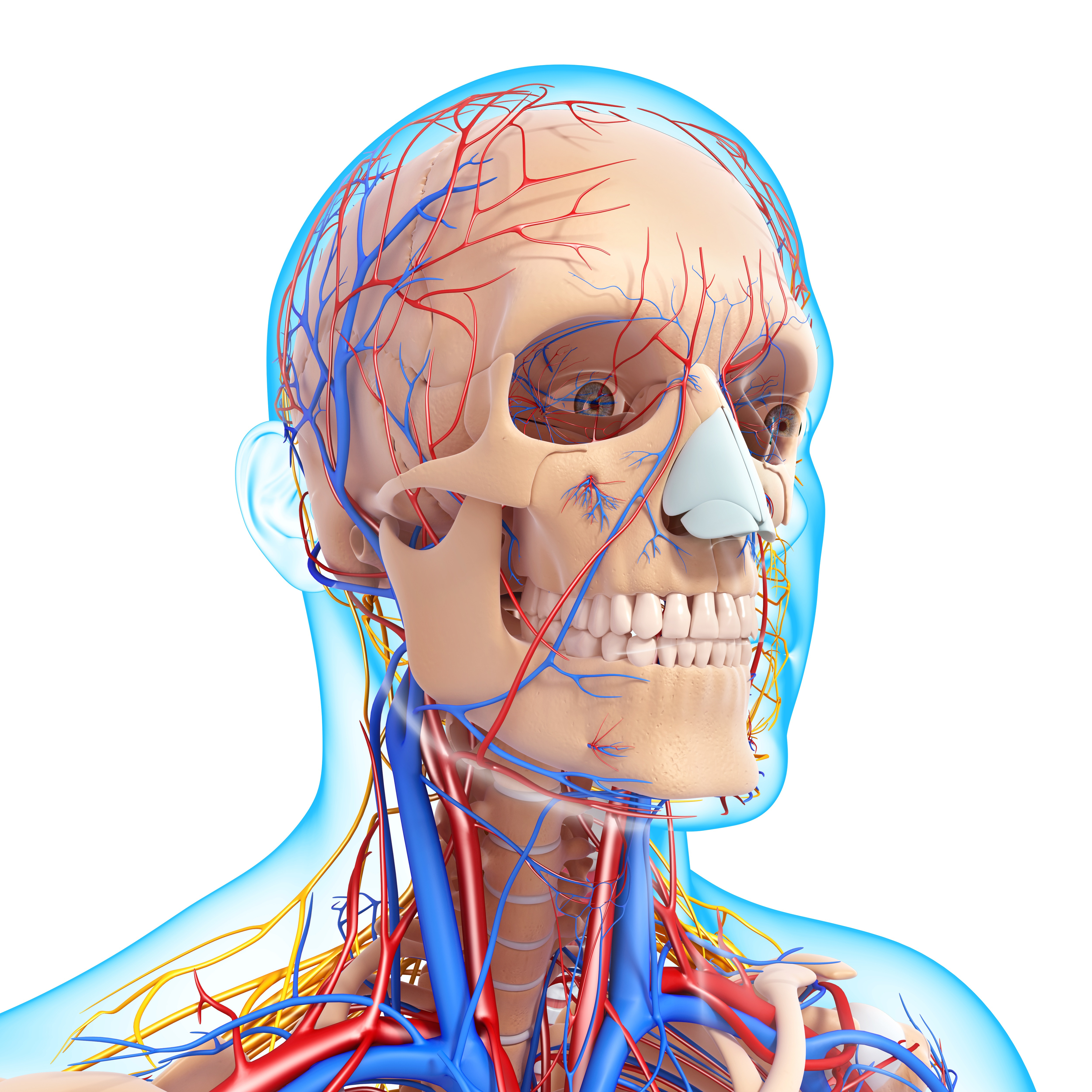
The orbital bones join to form the orbit or socket of the eye, where the eyeball rests.
The orbital structure provides pathways for the eye to connect with the nerves, lacrimal apparatus, adipose tissues, blood vessels, and extraocular muscles. This enables the eye to move and function properly.1 It also protects the eye from injury in case of head trauma.

However, these bones are delicate and can sustain fractures during a traumatic head injury, which may result from a car accident, impact during contact sport, or during a fistfight.
Can Orbital Bone Trauma Affect Vision?
Yes. A severe orbital bone fracture can cause vision issues such as double vision (diplopia), bruising around the affected eye, or difficulty making eye movements.
Other orbital fracture symptoms may include:
- Bruising due to blood pooling in areas around the eyes
- Eyeball changes, such as blood in the sclera (white part of the eye)
- Difficulty with or decreased eye movement
- Nausea or vomiting
- Sunken eyeballs
- Facial numbness due to nerve damage
- Eye or cheek pain
- Swelling due to inflammation of the forehead, cheek, or skin under the eye
Orbital Bone Anatomy & Functions
Seven types of bones form the different sections of the orbital structure. These include:
- Sphenoid
- Frontal
- Zygomatic
- Lacrimal
- Ethmoid
- Maxilla
- Palatine
Let's look at how these seven orbital bones join to form different parts of the eye socket (orbit):
- The orbital roof. Formed by the lesser wing of the sphenoid and the frontal bone.2
- The lateral wall. Formed by the greater wing of the sphenoid bone and the zygomatic bone.
- The medial orbital wall. Composed of the lacrimal, ethmoid, maxillary bones, and lesser wing of the sphenoid bones.
- The orbital floor. Consists of the palatine, maxillary, and zygomatic bones.3
The lateral wall is sturdier than the rest of the orbital walls. Together, the walls function as a barrier against trauma to the eye and anchor muscles that support eye movement. They also serve as a passage for nerves and blood vessels.
Types of Orbital Fractures
An orbital fracture means the bone is cracked or broken. There are three types of orbital fractures:
1. Orbital Rim Fracture
Orbital rim fractures are caused by trauma to the face, most commonly from a car accident. Considering the force required to cause this fracture, other injuries like facial bone and brain injury may also occur.
It may also cause damage to other parts of the eye, such as the optic nerve, eye muscles, tear ducts, and sinuses near the eye.
The two types of orbital rim fractures are:
- Zygomatic bone fracture. Occurs on the lower edge of the eye rim near the cheekbone.
- Frontal bone fracture (frontal sinus fracture). Occurs on the upper edge of the eye rim, near the forehead.
2. Direct Orbital Floor Fracture
Direct orbital floor fracture occurs when the rim fracture extends to the socket floor. The fracture is thought to occur due to increased intraorbital pressure. This causes orbital bones to break at their weakest point.
3. Blowout Fracture (Indirect Orbital Floor Fracture)
A blowout fracture occurs when the floor of the eye socket cracks. This causes a small hole that can interfere with eye muscles and other nearby structures. However, the bony rim of the eye is not affected.
Someone with a blowout fracture may experience difficulty moving their eyes, resulting in double vision.
This fracture may result from blunt impact with an object larger than the eye socket, such as a hockey puck, a baseball, a hammer, or a fist.
According to experts, about 85% of orbital bone fractures happen while playing a contact sport, doing repairs, at work, or during car crashes.4 Men who work in certain physically demanding jobs or have violent tendencies are at a greater risk of orbital fractures.5
Treatment Options for Orbital Fractures
An injured orbital bone requires immediate examination for any possible fractures. An experienced ophthalmologist can diagnose a bone fracture using X-rays or performing computed tomography (CT) scans.6
In most cases, the fracture can be treated with antibiotics, painkillers, decongestants, and cold compresses to reduce swelling.7 The body eventually heals on its own. However, in the case of a severe fracture, surgery may be the best option.
During recovery, bruises and swelling will heal first (within 7 to 10 days). The fracture may take longer to heal, and the timeline will depend on the severity of the damage.
Your ophthalmologist may advise against sneezing with the mouth closed, blowing your nose, or using a straw for several weeks to avoid further injury.
In this article
7 sources cited
Updated on February 20, 2024
Updated on February 20, 2024
About Our Contributors
Vincent Ayaga is a medical researcher and seasoned content writer with a bachelor's degree in Medical Microbiology. Specializing in disease investigation, prevention, and control, Vincent is dedicated to raising awareness about visual problems and the latest evidence-based solutions in ophthalmology. He strongly believes in the transformative power of ophthalmic education through research to inform and educate those seeking knowledge in eye health.
Dr. Melody Huang is an optometrist and freelance health writer with a passion for educating people about eye health. With her unique blend of clinical expertise and writing skills, Dr. Huang seeks to guide individuals towards healthier and happier lives. Her interests extend to Eastern medicine and integrative healthcare approaches. Outside of work, she enjoys exploring new skincare products, experimenting with food recipes, and spending time with her adopted cats.

