

The optic chiasm is the X-shaped crossing point where the optic nerves from each eye meet at the base of the brain.1 It splits and routes visual signals so each hemisphere of the brain receives information from both eyes, which is the structural basis for binocular vision and depth perception.1
When the optic chiasm is compressed or damaged, the result is usually a specific pattern of vision loss in both eyes rather than total blindness.6 This article walks through where the optic chiasm sits, what it does in the visual pathway, and the conditions that can affect it.
What Is the Optic Chiasm?
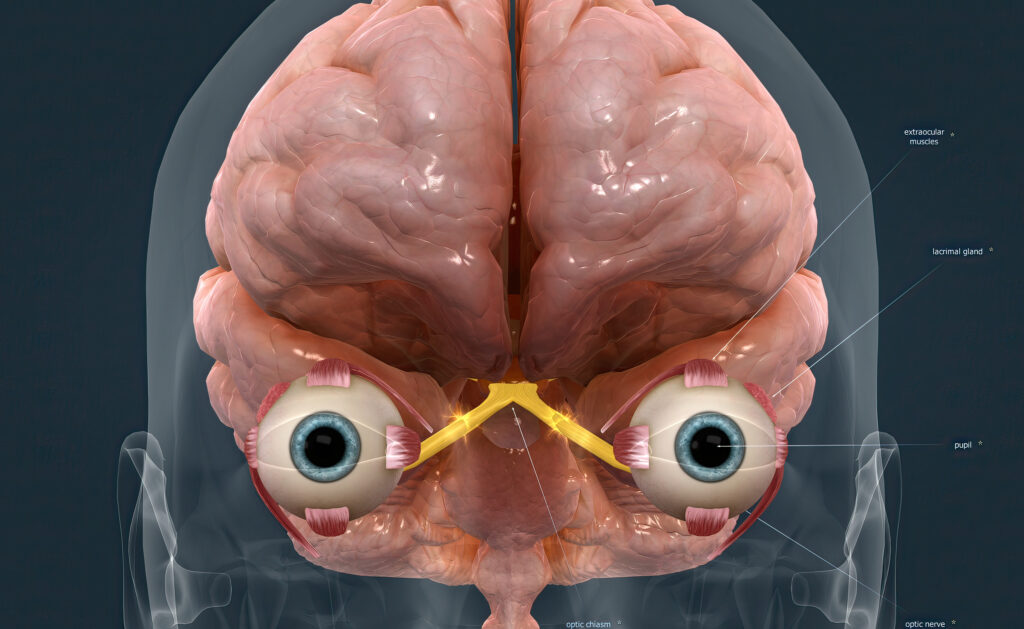
The optic chiasm, also called the optic chiasma, is an X-shaped structure at the base of the brain where the left and right optic nerves meet.1 Fibers from the inner (nasal) half of each retina cross to the opposite side, while fibers from the outer (temporal) half stay on the same side.1
That selective crossing is the whole point of the structure. It packages information from both eyes into two combined streams, one for the left visual field and one for the right, before sending them deeper into the brain.2
Where Is the Optic Chiasm Located?
The optic chiasm sits just above the pituitary gland and below the hypothalamus, in a region called the suprasellar cistern.1 Its proximity to the pituitary is what makes pituitary tumors a frequent cause of chiasm problems.6
The chiasm also lies in the anterior portion of the Circle of Willis, a ring of arteries that supplies blood to the brain.3 The Circle of Willis is one of the most common sites for intracranial aneurysms, which is why vascular conditions can sometimes involve the chiasm.3
What Does the Optic Chiasm Do?
The optic chiasm sorts and routes visual signals so each side of the brain processes the opposite side of your view.2 Nasal retinal fibers cross to the opposite side while temporal fibers stay ipsilateral, so the right optic tract carries signals from the left visual field and the left optic tract carries signals from the right visual field.2
This routing is what enables organized binocular visual processing.1 Without it, your brain could not fuse the slightly different images from your two eyes into a single, depth-rich view of the world.10
The optic chiasm supports:
- Binocular vision (using both eyes together)10
- Stereopsis (depth perception)
- The ability to judge distance accurately
- Visually guided hand movement and hand-eye coordination
- A unified view across your full visual field
Visual Pathway of the Optic Nerve
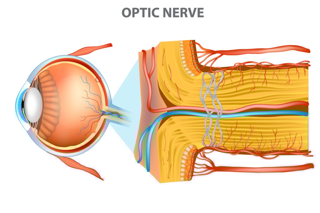
The visual pathway is the route light signals take from your retina to the visual cortex at the back of your brain.11 The optic nerve, also called the second cranial nerve or CN II, is the first leg of that journey.4
Each optic nerve starts in the retina, the light-sensing tissue at the back of the eye, and ends at the occipital lobe of the brain.11 The pathway runs through these structures:
- Optic nerve fibers pick up signals from retinal cells
- Nerve fibers pass through the optic canal to reach the middle cranial fossa4
- Fibers from each eye meet at the optic chiasm, where nasal fibers cross to the opposite side
- Reorganized fibers enter the left and right optic tracts
- Each tract travels to the lateral geniculate nucleus of the thalamus
- Fibers synapse and continue as the optic radiations to the visual cortex
- The visual cortex interprets the signals as the images you see2
What Happens If the Optic Chiasm Is Damaged?
Damage to the optic chiasm typically produces a distinctive pattern of vision loss in both eyes at once, most often bitemporal hemianopsia (loss of the outer half of vision in each eye).5 The exact pattern depends on which fibers are affected and whether the lesion sits at the center, edge, or junction with the optic nerve.6
Visual field patterns linked to chiasmal or surrounding pathway damage include:
- Bitemporal hemianopsia: loss of the outer (temporal) peripheral vision in each eye, the classic chiasmal sign5
- Junctional scotoma: a blind spot caused by a lesion at the junction of the optic nerve and chiasm, affecting one eye more than the other6
- Homonymous hemianopsia or quadrantanopia: loss of the same side or quadrant of vision in both eyes, usually pointing to damage behind the chiasm in the optic tract or radiations6
Conditions that can involve the optic chiasm include:
- Pituitary adenomas and other compressive tumors (the most common cause)6
- Vascular disorders, including aneurysms of the Circle of Willis3
- Inflammatory and demyelinating disease, such as multiple sclerosis9
- Infectious causes, such as tuberculosis or viral chiasmitis (uncommon)9
- Traumatic head injury near the skull base1
Untreated compression can cause progressive visual dysfunction and, if it continues, may lead to irreversible optic nerve damage.8 That is why steady changes in side vision warrant prompt evaluation rather than a wait-and-see approach.8
Pituitary Adenomas
A pituitary adenoma is a tumor of the pituitary gland that sits directly below the optic chiasm.7 Most are benign, but larger tumors (macroadenomas) can grow upward and press on the chiasm from below.7
When a pituitary tumor compresses the chiasm, it typically affects the crossing nasal fibers first, which is why bitemporal vision loss is the hallmark presentation.6 Treatment can include medication, surgery, or radiation depending on the tumor type and size.7
When to See Your Doctor
Sudden or progressive vision changes that involve both eyes deserve same-day medical attention.12 The patterns most associated with optic chiasm or visual pathway problems are not subtle once they take hold, but early signs can be easy to dismiss.
Seek prompt evaluation if you notice:
- Sudden partial or complete vision loss in one or both eyes12
- A curtain or shade moving across your field of view12
- New blind spots, especially in the outer halves of your vision12
- Persistent double vision or sudden distorted vision12
- Painful blurred vision or vision loss with severe headache12
An optometrist or ophthalmologist can run a visual field test that maps exactly where your vision is intact and where it is not.6 If results suggest a lesion at or near the chiasm, imaging of the brain and pituitary is the next step.6
Summary
The optic chiasm is the X-shaped junction at the base of the brain where the optic nerves from each eye meet and partially cross.1 Its job is to route visual information so each side of the brain handles the opposite side of your field of view, which is the foundation of binocular vision and depth perception.1
Because the chiasm sits right above the pituitary gland and inside the Circle of Willis, it is vulnerable to compression from pituitary tumors, vascular conditions, and inflammatory disease.6 The classic sign of chiasm involvement is bitemporal hemianopsia, a loss of side vision in both eyes that should always trigger a prompt eye and neurological work-up.5

