What Are the Different Parts of the Eye?
The human eye is a complex organ composed of several interconnected parts, each with a specific function in vision. Let's explore these components and their roles in enabling us to see the world around us.
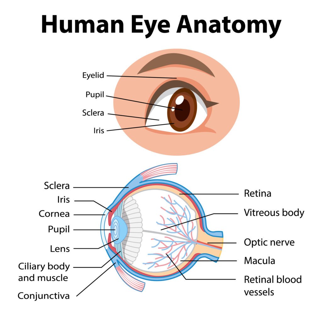
The following are parts of the human eyes and their functions:
1. Conjunctiva
The conjunctiva is the membrane covering the sclera (white portion of your eye). The conjunctiva also covers the interior of your eyelids.
The conjunctiva helps lubricate the eyes by generating mucus and tears. It also aids in immunological monitoring and prevents microorganisms from entering the eye.
Pink eye (conjunctivitis) occurs when this thin membrane becomes inflamed or swollen. Other eye disorders that affect the conjunctiva include:
- Pinguecula. Accumulation of protein and fat deposits in the conjunctiva.
- Pterygium. A noncancerous growth that develops on the conjunctiva.
- Subconjunctival hemorrhages. broken blood vessels.
2. Sclera
The whites of the eye (sclera) cover more than 80% of the eyeball’s surface. The sclera has a smooth, white exterior but is brown on the inside.
It has grooves that help properly attach the eye tendons, providing stability and protection while staying flexible. This allows the eye to move as needed to see different objects.
The episclera is a thin layer of tissue on top of the sclera that has tiny blood vessels that provide the sclera with nutrients. If the sclera or episclera becomes inflamed, it results in a condition known as scleritis or episcleritis, respectively. These conditions can cause:
- Redness
- Eye pain
- Blurry vision
- Lid swelling
3. Iris
The iris is the colored part of the eye and is unique to each person. This structure is located in the front of the eye, between the cornea on the outside and the lens on the inside.
The iris primarily regulates how much light reaches the retina by controlling the size of the eye’s “window,” or pupil. As a result, it narrows in bright light while opening up in low light. This is also referred to as the pupillary light reflex.
The iris also performs what is known as the “accommodation reflex”, which is the eye’s instinctive ability to shift focus from nearby to distant objects. This action requires adjusting the pupil’s aperture (opening), the shape of the lens, and convergence (the ability of the eyes to work together).
4. Pupils
The pupil is seen as a black dot in the center of the iris. It’s essentially a hole that allows the eye to focus on the things in front of it. Similar to the iris, they open and close to regulate the amount of light that enters the eye.
When light enters the eye through the lens, it focuses light rays through the pupils and into the retina. The difference between the centers of your pupils is called your pupillary distance.
When it’s dark, our pupils dilate or expand wider to let in more light, increasing the scope of our view. In bright light, our pupils contract to a small diameter to protect our retina’s delicate photoreceptors.
5. Cornea
The cornea is the clear and protective outer layer of your eye. Along with the sclera, the cornea is a barrier against dirt, infectious microorganisms, and other substances that can damage the eye.
Aside from protection, the cornea also plays a significant role in vision. Its dome-shaped surface bends light as it passes through the eyes, allowing it to focus on objects effectively.
The cornea can also filter out the sun’s harmful ultraviolet (UV) light, preventing it from reaching other structures inside the eye. However, you should still wear sunglasses to protect your eyes. Chronic exposure to UV light may lead to inflammation and other complications, including cancer.
6. Uvea
The uvea is the eye’s middle layer. It is located underneath the white part of the eye (the sclera). It’s composed of three parts, namely, the iris, the ciliary body, and the choroid.
These structures control some eye functions, such as adapting to varying levels of light or object distances. If any structures become inflamed, the resulting condition is called uveitis.
7. Choroid
This vascular layer is located between the sclera and retina of your eye. It delivers nourishment from the blood and oxygen supply to the retina’s outer layers.
Essentially, the choroid is the source of life that keeps the retina functioning effectively. It also reflects light, causing the red-eye effect in photographs.
8. Retina
The retina is a light-sensitive layer that covers your eye’s rear surface. Images are transmitted to the retina when your eye picks up the images.
It converts images into impulses that are sent to your brain through the optic nerve, allowing you to see and interpret images. Some of the ocular conditions that affect the retina include:
- Diabetic retinopathy. Diabetes complications caused by damaged blood vessels.
- Retinal detachment. When the retina detaches from its normal position.
- Retinitis pigmentosa. Deterioration of special light-sensitive cells in the retina.
- Retinoblastoma. Formation of cancer cells in the retinal tissues.
9. Eye Muscles
The eye has six muscles that come from the eye socket (orbit) and work to move the eye up and down, side to side, or in a circular motion. These muscles include:
- The superior rectus. Attaches to the top of the eye and moves the eye upwards.
- The inferior rectus. Attaches to the bottom of the eye and allows downward eye movement.
- The medial rectus. Attaches to the side of the eye adjacent to the nose and helps the eyes to shift inwards towards the nose.
- The lateral rectus. Attaches to the outer side of the eyes and moves the eyes toward the temples.
- The superior oblique. Originates from the back of the eye socket and attaches to the top of the eye. It rotates the eye inwards (front to back) and downwards.
- The inferior oblique. Arises from the front of the socket near the nose and travels inwards, attaching to the bottom surface of the eyeball.
10. Macula Lutea
Light rays are focused on the macula lutea when an eye looks directly at an object. The macula lutea is a yellow oval area in the retina’s center (back of the eye). The center of the macula is known as the fovea.
The macula lutea is the section of the retina responsible for sharp, detailed central vision (visual acuity). It has a high concentration of cones, which are the light-sensitive retinal cells that provide high visual acuity.
11. Eye Lens
The lens of the eye is the transparent lentil-shaped structure inside your eye. It’s a natural lens located behind the iris and to the front of the vitreous humor which is a clear, colorless, gelatinous mass that fills the gap between the lens and the retina.
The lens is held in place by a fibrous membrane known as the zonule of Zinn or the lens suspensory ligaments. The lens changes its thickness and curvature, allowing the eye to focus on objects from varying distances.
If your lens has an irregular curvature, you’re more prone to develop astigmatism. Cataracts are another lens-related visual disorder in which the lens becomes opaque or hazy, impairing vision.
12. Aqueous Humor
Aqueous humor is a fluid substance that fills the eye. It’s divided into two chambers. The anterior chamber is in front of the iris, whereas the posterior chamber is right behind it.
These layers enable the eye to keep its shape. The aqueous fluid is evacuated via the Schlemm canal to eliminate any accumulation in the eye. If a person’s aqueous fluid does not drain adequately, glaucoma may develop.
13. Ciliary Body
The ciliary body is a ring-shaped tissue found behind the iris. It attaches to the lens through the zonular fibers (fibers of Zinn).
The ciliary body holds and regulates the eye lens’s movement, keeping the lens shape intact. This structure is also involved in the production of aqueous humor.
14. Optic Nerve
The optic nerve is a bundle of about 1.2 million nerve fibers that transmit visual information to the central nervous system (CNS). There is one nerve per eye connecting each eye to the brain.
Vision loss may occur if any of the nerves are damaged. However, the consequences of optic nerve damage depend on the extent of the damage.
15. Optic Disc
The optic disc is where the axons of retinal ganglion cells join together and mark the beginning of the optic nerve (second cranial nerve). It also serves as the entrance site for major blood vessels that nourish the retina. In the average person, the optic disc carries about 1.2 million nerve fibers from the retina to the brain.
16. Fovea Centralis
The fovea centralis (central fovea) is a tiny depression in the retina that houses cones that help with proper vision. It is located within the macula. Approximately 80-90% of the optic nerve fibers convey visual information from the fovea.
Meanwhile, the other portion conveys information from the peripheral retina. If the fovea of the cones is affected, you may experience blurry vision.
Common Eye Conditions That Can Affect Vision
Various eye conditions can impact how well you see, these include:
The best way to prevent these problems is to maintain your physical health, get regular eye exams, and eat a healthy balanced diet.