Iris Anatomy and Functions
The iris is the colored part of the human eye and a component of the uvea (uveal layer or uvea coat). The uvea is a pigmented layer found between the retina and the sclera (white of the eye).
In addition to the iris, the uvea also consists of the choroid and ciliary body. The choroid is a vascular layer found between the retina and the sclera. It supplies blood and oxygen to other parts of the retina.
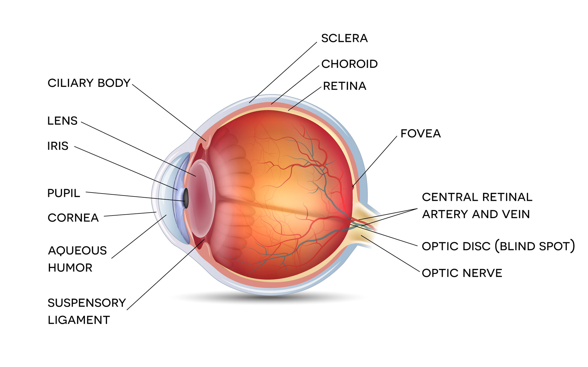
The ciliary body is located behind the iris. It consists of muscles that shape the lens when the eye focuses. It also makes the aqueous humor, which is a clear fluid in the space between your cornea and iris.
Within the iris is an opening called the pupil. This is a passage for light as it enters the eye. The iris controls pupil size and the amount of light getting into the eye.
Pupil Size
A normal adult pupil ranges from 2 to 4 millimeters in diameter in bright light. In the darkness, the diameter may range from 4 to 8 millimeters. The pupil’s size may change due to other reasons such as:
- Your emotions (e.g., happiness or sadness)
- Underlying conditions (e.g., headache or double vision)
- Drug abuse (e.g., cocaine, heroin, and LSD)
The iris has two types of smooth muscles that change the size of your pupils, sphincter pupillae and dilator pupillae. The sphincter pupillae make the pupil contract, whereas the dilator pupillae enlarge them. The sympathetic and parasympathetic nervous systems, which send signals to the muscles to contract or expand, also make this process possible.
Eye Color
Everyone, regardless of their eye color, has the same type of melanin pigment. Melanin is a natural skin color produced by cells known as melanocytes. Melanocytes are found in hair, skin, and the iris of your eyes.
Everyone has the same number of melanocytes. However, they produce different amounts of melanin, which causes different eye colors.
The color of your iris depends on the amount of melanin pigment in your iris. Sufficient amounts of melanin will make your iris brown. A reduced amount of melanin allows light to pass through and get scattered by collagen fibers, resulting in a bluer color.
Depending on the extent of pigmentation, the color can range from light blue to dark brown. Common eye colors include blue, green, hazel, or brown. Nowadays, iris color is an inherited trait.
Accommodation Reflex
The iris regulates the light entering the eye by changing the size of the pupil based on the situation. For example, when it’s dark, the iris muscles expand the pupils to allow more light into the eye. Similarly, it contracts to reduce the amount of light entering the eye when in bright light.
The iris muscles enable the eye to shift focus based on near or far objects. The ciliary muscle changes the shape of the eye lens to help you see clearly.
Common Iris and Pupil Disorders
Below are common eye conditions that affect the iris and pupil:
- Narrow-angle glaucoma. Causes the iris to bulge forward, blocking or narrowing the cornea-iris angle. It also affects eye fluid movement, resulting in increased eye pressure.
- Iris melanoma. This is cancer of the iris. The condition usually has no symptoms, but the tumor may be noticeable during a routine eye exam.
- Iritis. An infection that develops in one or both eyes. It’s characterized by red, painful eyes accompanied by a headache, blurred vision, and light sensitivity.
- Horner syndrome. Occurs when the sympathetic nerves are damaged by physical injury, tumors, or other diseases. It can cause permanent pupil contraction.
- Iris coloboma. A hole in the iris caused by eye trauma or after eye surgery. It can also be inherited.
These conditions often require medical attention. For example, narrow-angle glaucoma is considered a medical emergency. Meanwhile, iritis can lead to glaucoma, cataracts, and vision loss.
Harmless Eye Conditions
One of the more common eye conditions that are harmless is anisocoria. It’s a condition that causes your pupils to appear in different sizes.
The condition is genetic, but different family members may have pupils of different sizes. Additionally, it may result from an underlying condition such as Horner’s syndrome or other diseases of the brain, nerves, and blood vessels.
Another harmless condition is heterochromia, which occurs when a person has two different-colored eyes. It can be either complete or partial heterochromia.
Complete heterochromia occurs when one iris is different from the other, and partial heterochromia occurs when a part of one iris is a different color from the other.
Treatment for Iris and Pupil Problems
Routine eye exams are essential for protecting your eyesight and preventing issues from becoming worse. Your doctor can even provide medical advice on eye care.
Treatment options for iris disorders include:
- Artificial iris. When you have a dysfunctional iris, you can get an implant. For example, CustomFlex Artificial Iris was approved in 2018 for treating congenital aniridia.
- Steroid eye drops. These are often used to reduce inflammation.
- Dilating eye drops. These are prescribed to dilate your pupil and reduce eye pain, such as in the case of iritis.
- Antibiotic or antiviral eye drops. These are prescribed to prevent infection, especially after eye trauma.
- Contact lenses. If your iris problem causes poor vision, your doctor may prescribe contact lenses. Prosthetic-colored contact lenses can also hide iris defects and improve visual symptoms like glare.
- Laser surgery. Sometimes, your eye doctor may suggest surgery to prevent damage caused by some conditions. Laser Peripheral Iridotomy (LPI) is used to treat narrow-angle glaucoma.