The retina is one of many vital parts of the human eye that enable you to see. This nerve layer at the back of the eye contains light-sensitive cells called rods and cones. The retina senses light and generates electrical impulses so the brain can create an image.
This article discusses the retina’s anatomy, functions, and associated disorders. It also covers the fundamental anatomy of the eye and how the retina works with other primary structures.
Anatomy of the Retina
The human retina consists of layers of neural tissue that line the entire back wall of the eye. It’s the only extension of the brain visible from outside the body (via a retinal exam).1
The retina attaches to the optic nerve at the optic disc. The optic nerve is one of the main cranial nerves coming from the brain.
Many parts of the eye are involved with the retina. These include:
Photoreceptors
The retina contains millions of light-sensitive cells called photoreceptors. These specialized cells are responsible for turning light into signals that are sent to the brain.
There are two main types of photoreceptors:
- Rod cells. Also called rods, these cells are extremely sensitive to light. They help you see in dim light conditions.
- Cone cells. Cones provide color vision and help you see fine details. There are three types of cone cells: red, green, and blue. They function best in bright light.
Macula
The macula is the retina's center, where cone cells are concentrated. This part of the eye helps you see details in the center of your visual field.
Age-related macular degeneration is a common condition when this area of the retina deteriorates.
Fovea
The fovea is the very center of the macula, where vision is the sharpest. The high concentration of cone cells provides acute vision by creating a sharp, accurate image.
Peripheral Retina
This is the area of the retina outside the macula, responsible for peripheral vision and night vision. Rod cells are more abundant in the peripheral retina.
Functions of the Retina
The retina is responsible for converting light entering your eye into visible images. It’s often compared to film in a camera.
Even though vision is commonly associated with the eyes, the brain turns visual information into recognizable images. The retina acts as a bridge between the human eye and the brain. It’s where light rays become signals your brain interprets as images.
The retina creates electrical signals when light rays enter the eye through the pupil. These signals travel through the optic nerve, where the brain processes them in the visual cortex.
Visual processing in the brain enables you to perceive the shape, design, color, and other fine details of the objects you look at.
Basic Anatomy of the Eye
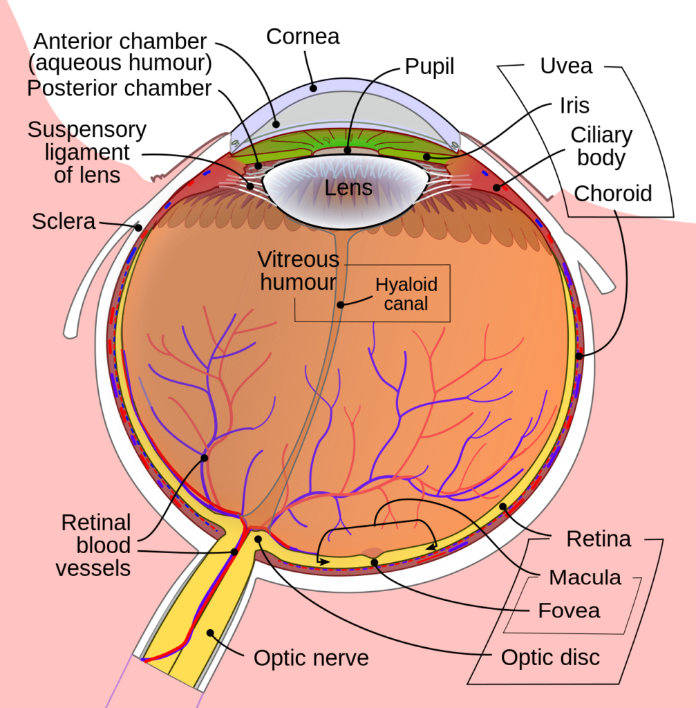
The human eye is a highly complex organ with many essential parts. Aside from the retina, which is explained in detail below, key parts of the eye include:
- The cornea. The clear dome at the front of the eye covering the iris and the pupil. Light passes through the cornea and lens, which bend (refract) it to focus on the retina.
- Pupil. The black dot at the front and center of the eye is an opening through which light enters.
- Iris. The colored part surrounding the pupil which helps control how much light enters the eye. The iris makes the pupil smaller in bright light and larger in dim light.
- The lens. This transparent structure inside the eye helps refract incoming light onto the retina. The lens and the cornea both work to provide clear vision.
- Sclera. The white part of the eye.
- Vitreous humor. This transparent gel-like fluid inside the eye helps it maintain a round shape.
- Optic nerve. This bundle of nerve fibers connects the retina to the brain, where all the visual information is processed.
Vision Problems and Disorders Associated with the Retina
Retinal conditions vary widely and can affect any part of your retina. Most retinal diseases cause vision problems.
Common retinal problems include:
Diabetic Retinopathy
Uncontrolled diabetes can cause the tiny blood vessels at the back of the eye to weaken and leak into the retina. Diabetic retinopathy can lead to vision loss and blindness.
Age-Related Macular Degeneration
Deterioration of the retina's center (macula) is common in people over 50. Age-related macular degeneration (AMD) affects central vision and your ability to make out details.
AMD may cause a blurry, distorted view or a blind spot at the center of your visual field. Over time, this can lead to vision loss.
Retinal Tear
This occurs when there’s a rip or tear in the retina. Without treatment, a retinal tear may lead to retinal detachment, a medical emergency.
Symptoms of a torn retina include sudden blurry vision, increased eye floaters, and flashes of light.
Retinal Detachment
This occurs when the retina detaches from other parts of the eye that provide nourishment and function.
A retinal detachment is considered a medical emergency. Seek immediate medical care if you experience a sudden increase in eye floaters, light flashes, or a shadow in your visual field.
Macular Hole
A macular hole is a small hole in the macula. The hole may occur on its own or develop due to an eye injury. It usually interferes with your central vision.
Macular Pucker
A macular pucker occurs when scar tissue causes the retina to wrinkle. A pucker can result from a retinal tear, detachment, or other retinal condition. It may or may not affect your vision.
Retinitis Pigmentosa
This is a rare genetic disorder that involves the loss of retinal cells.6 Retinitis pigmentosa causes night blindness and loss of side vision (peripheral vision).
Posterior Vitreous Detachment (PVD)
This condition occurs when the gel that fills the inside of the eye (vitreous humor) separates from the retina. Symptoms of PVD include increased floaters and flashes of light.
PVD may lead to a retinal tear, detachment, macular hole, or pucker.
Why Routine Eye Exams are Important
During routine eye examinations, your eye doctor examines your retinas for signs of diseases. They may be able to detect early signs of retinal conditions before you even notice vision changes.
Regular examinations enable your doctor to detect any issues early before they become serious. Most eye issues are treatable during the early stages.
In addition to routine exams, call your eye doctor immediately if you have sudden changes to your vision. This includes blurry vision, increased floaters, and flashes of light.
Summary
The retina plays an essential role in the anatomy of the eye. It’s the light-sensitive layer covering the back of the eye.
The retina includes light-sensitive cells that convert incoming light into electrical signals. These signals pass through the optic nerve to the brain, which processes them into images.
It’s crucial to see your eye doctor for routine retinal exams.