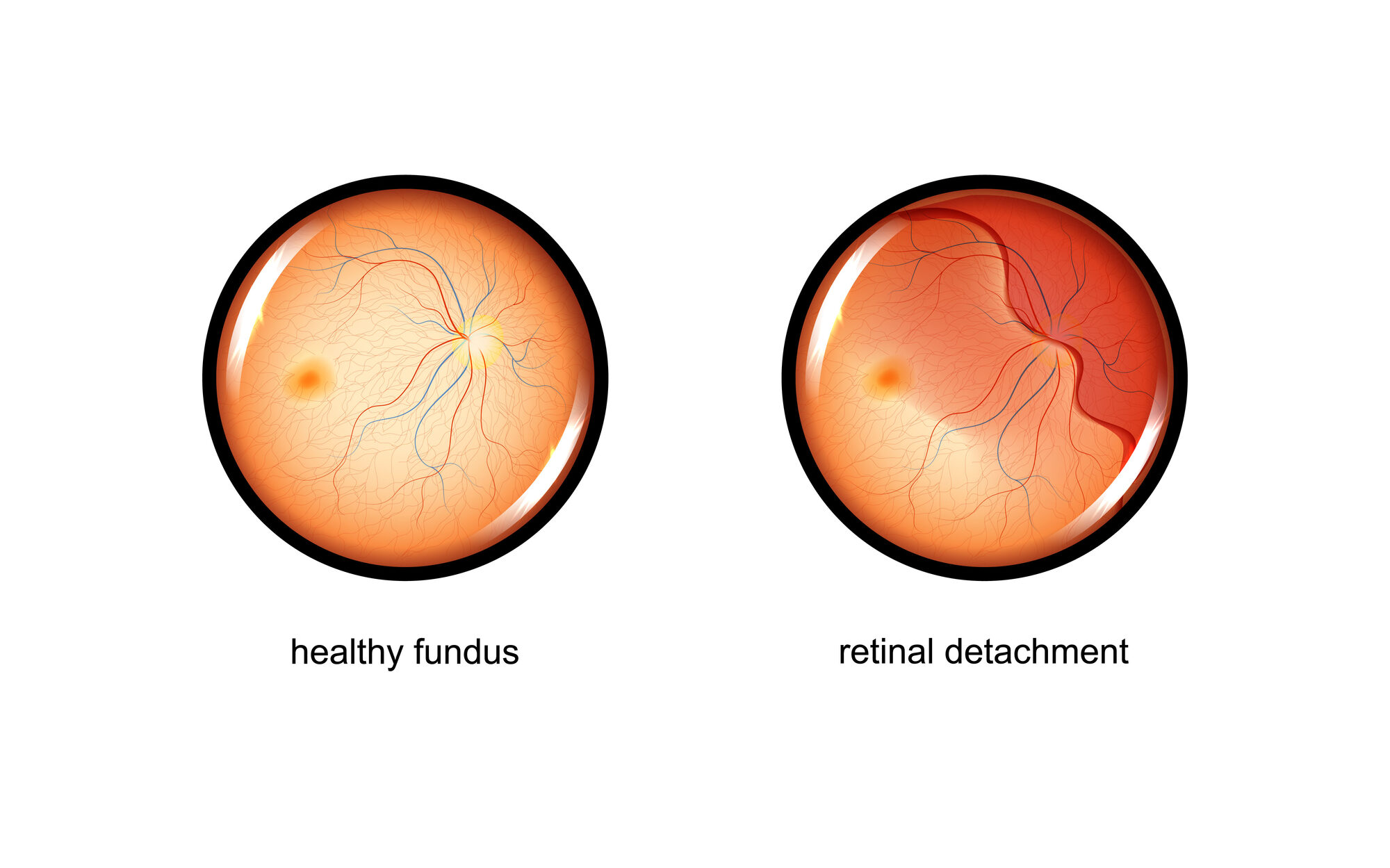
A retinal tear occurs when the retina develops a small break or hole, allowing fluid from the vitreous gel inside your eye to seep underneath. This can potentially lift the retina away from its normal position, resulting in retinal detachment.
A detached retina can seriously harm your vision, making rapid action essential. Because of this, it’s important to understand the signs and symptoms of a retinal tear.
In this article, we’ll discuss everything you need to know about retinal tears and when to get help. If you have a retinal tear, you may need treatment to prevent retinal detachment.
What Causes Retinal Tears?
As you age, the vitreous gel filling your eye naturally shrinks and becomes more watery, eventually pulling away from the retina. This process, called posterior vitreous detachment (PVD), can tug hard enough to tear the retina in one or more places.
A retinal tear can happen after an injury or through age-related vision changes. However, certain people have a higher chance of experiencing a retinal tear:
- High myopia (nearsightedness)
- Lattice degeneration
- Previous eye surgery (especially cataract surgery)
- Family history of retinal detachment
When Should You Call a Doctor?
Early detection and immediate care can significantly increase the likelihood of preserving your vision. Contact your eye doctor right away if you notice:
- Flashes of light, like quick sparks or lightning streaks, especially at the edges of your vision
- Sudden eye floaters or increased dark specks drifting through your vision
- A dark shadow or curtain appearing in your peripheral (side) vision, slowly progressing to your central vision
- Sudden blurry vision
How Are Retinal Tears Treated?
Your doctor will recommend treatment based on the size, location, and severity of your retinal tear or detachment. Here are the common approaches, explained in straightforward terms.
For small, recent tears without detachment, your retina specialist may quickly seal the tear in an office procedure:
- Laser photocoagulation. Tiny laser burns create scar tissue, sealing the edges of the tear.
- Cryopexy (freezing treatment). A freezing probe applied outside your eye creates a similar seal.
These procedures have excellent success rates, typically above 90%, with minimal downtime. You can usually return to normal activities within a few days to several weeks, depending on your surgeon’s instructions and the specific procedure.
Surgical Repairs for a Detachment
Larger tears or detachments usually require surgery. Your doctor chooses the procedure based on your specific needs:
- Pneumatic retinopexy. A gas bubble injected into your eye pushes the retina back into place, combined with laser treatment. It is often office-based with a fast recovery, but is best suited to smaller detachments.
- Scleral buckle. A flexible silicone band wraps around your eye to relieve pulling forces. It's often recommended for younger or highly myopic patients.
- Pars plana vitrectomy. This is the most common and versatile option. The surgeon removes the vitreous gel, replaces it with a gas bubble or silicone oil, and seals the retina with laser or cryotherapy.
Combined procedures may be performed depending on your retina's condition. Your surgeon will carefully choose the best option to restore your vision effectively.
Conservative Treatments
Not all retinal tears require aggressive intervention. These low-risk asymptomatic breaks (such as atrophic holes or operculated breaks) may not require immediate treatment.
For these cases, careful monitoring is essential. Sometimes the eye spontaneously starts to form a stabilizing scar around the tear.
Once a tear has been identified, it must be followed by a trained specialist to ensure it doesn’t worsen. Retinal tears need to be continually monitored by a specialist who can take action if necessary.
What To Do After Surgery
Recovery varies by procedure, but clear guidelines help you plan your life after retinal surgery:
- Avoid driving or working for a few days until your surgeon clears you.
- Avoid intense exercise and heavy lifting.
- Avoid dusty environments.
- Avoid touching or rubbing your eyes, especially with dirty hands.
- Avoid getting water directly onto your eyes.
- Don’t wear eye makeup for several weeks, and avoid contact lenses until your surgeon says it’s safe.
You may need specific head positioning for days (often up to a week or more) to help the retina heal properly. Apps and specialized pillows make this easier.
How Does A Retinal Tear Lead to Detachment?
When the retina tears, vitreous fluid can slowly pass through the break, creating a layer of fluid beneath the retina. Gradually, this fluid pushes the retina away from the eye’s inner wall.
As this progresses, you may notice a curtain-like shadow descending or rising into your line of sight, signaling the start of a detachment.
The outcome for a retinal tear is generally good as long as you receive treatment early and before the central retina (macula) detaches. Keeping track of symptoms can help your specialist provide timely treatment.
How Is a Retinal Tear Diagnosed?
An accurate diagnosis helps your doctor determine if immediate laser treatment or surgery is needed. Your eye doctor will perform a thorough exam by dilating your pupils with special drops.
This dilated eye exam allows a clear view of your retina and helps detect tears or detachment. Your doctor may also gently press on your eye for scleral depression to inspect peripheral areas.
If bleeding or cloudy fluid inside your eye obstructs your doctor's view, your doctor might use an ultrasound to evaluate the retina. Some clinics may perform additional imaging, such as optical coherence tomography (OCT), to further clarify your condition and guide the next steps.
Listen In Q&A Format
Retinal Tear Causes Symptoms Treatments
Vision Center Podcast
Prevention and Future Eye Safety
While not all risks can be eliminated, you can take meaningful steps to reduce your chances of future retinal issues:
- Wear protective eyewear during sports or activities with eye injury risks.
- Promptly report symptoms in your fellow eye, such as new floaters or flashes.
- Keep regular follow-ups with your eye care provider, especially if you've had retinal issues previously.
- Avoid flying, high altitude, or scuba diving while a gas bubble is in the eye; resume only after your surgeon confirms the gas is fully gone.