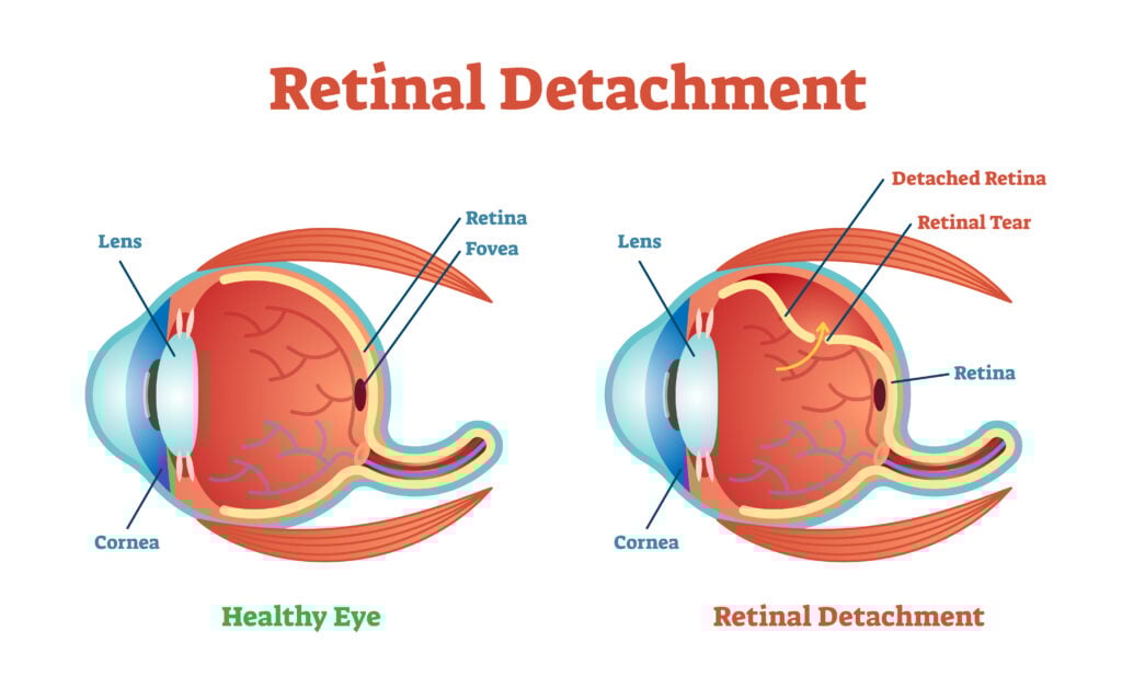
Scleral buckle surgery, or scleral buckling, repairs a retinal detachment. The retina is a layer of tissue located inside the eye. It sends visual information from the optic nerve to your brain.
It’s a medical emergency that requires immediate attention and care by a doctor. If left untreated, retinal detachment can lead to permanent vision loss.
During the procedure, a surgeon attaches a piece of silicone or a sponge to the white of the eye (sclera). The buckle helps repair retinal detachment by pushing the sclera toward the retinal break or tear.
When is Scleral Buckling Necessary?
Scleral buckle treatment is used for different types of retinal detachments. These include:
- Rhegmatogenous retinal detachment
- Inferior retinal detachments
- Complex detachments with multiple retinal tears
- Retinal detachment in young patients who haven’t undergone cataract surgery
- Recurrent retinal detachment
How Does a Scleral Buckle Procedure Work?
Scleral buckling is typically an outpatient procedure performed by an eye surgeon or retina specialist. The surgical procedure takes about 60 to 90 minutes.
How to Prepare for Scleral Buckling
Before scleral buckling, your eye doctor will inform you of any medications or foods you must stop taking beforehand.
You’ll also have to arrange for someone to pick you up after the surgical procedure.
What Happens During a Scleral Buckling Procedure?
Here are the procedure steps you can expect during eye surgery:5
- Your surgeon will numb your eye with local anesthesia. You might also receive general anesthesia before the surgery to fall asleep.
- You will receive eye drops to dilate your eyes.
- Your surgeon will make an incision into the outer layer of your eye (sclera).
- To stop a detachment or retinal tear from reopening, your doctor may also perform laser photocoagulation or cryopexy. This creates scar tissue to seal the break.
- A buckle or sponge is stitched around the sclera.
- Following surgery, the surgeon will drain any fluid behind your retina and apply antibiotic eye drops to prevent infection.
Scleral buckling is typically permanent. If you have a minor retinal detachment, the doctor might place a temporary buckle that will be removed once your eye heals.
How Much Does a Scleral Buckle Procedure Cost?
Scleral buckling typically costs $3,000 to $5,000, including surgical costs, anesthesia fees, and hospital charges.
Fortunately, most health insurance plans cover at least part of the cost because retinal detachment is a medical emergency. Talk with your doctor and/or insurance provider to determine your options.
Vision Results After Scleral Buckle Surgery
Scleral buckle surgery is successful in about 90% of cases.4 The chances for clear vision following surgery are higher if the macula was still attached before surgery.
If your detachment affected the macula, good vision following scleral buckling is still possible but less likely. You should have a follow-up eye exam about six months after surgery to check for vision changes.
Potential Complications and Scleral Buckling Risks
Overall, scleral buckle surgery produces positive results. However, every surgery involves some level of risk.
Possible complications of scleral buckle surgery include:
- Infection
- Double vision (diplopia)
- Cataracts
- Bleeding in the eye
- Glaucoma (increased intraocular pressure)
- Another retinal tear or detachment
- Becoming more nearsighted
If you’ve had previous eye surgery and existing scar tissue, this surgery might not immediately repair a retinal detachment.
If a repeat surgery is necessary, your eye doctor will remove existing scar tissue before proceeding. Scar or pre-existing scar tissue can affect the retina’s ability to reattach.
Scleral Buckle Recovery Time and Aftercare
Recovery time from scleral buckle surgery is anywhere from two to four weeks.
Your doctor will give you aftercare instructions following surgery. This includes information on when you can start taking prescription medications and instructions for taking post-surgery pain medications.
Days 1 to 2
You can typically return home on the day of surgery, but you will need someone to drive you.
You should expect some temporary side effects, including:
- Redness and swelling
- Tenderness
- Minor pain and discomfort
You must wear an eye patch for a couple of days following surgery. You'll also apply antibiotic eye drops to prevent infection. You must apply eye drops for up to six weeks following surgery.
Days 2 to 3
Swelling can occur after scleral buckling surgery. Your doctor might recommend placing an ice or cold pack over the eye for 10 to 20 minutes to lessen swelling. Wrap the ice pack around a clean towel before setting it on your skin.
Some doctors suggest applying an ice pack during the first three days following surgery, every one to two hours.
Days 3 to 14
Wait for your eye to heal before engaging in strenuous activity. During this period, avoid exercise, cleaning, and heavy lifting.
Your doctor may also suggest limiting the amount of reading to prevent too much eye movement.
Weeks 2 to 5
Some people can go back to work after two weeks. But this depends on how you feel and the nature of your work.
You should stay at home longer if your role involves heavy lifting or a lot of computer work.
Weeks 6 to 8
Meet with your doctor for a follow-up eye exam. The doctor will check the surgical spot to monitor your healing progress.
They will also assess if there is any improvement in sight. They may recommend corrective lenses or a new eyeglass prescription.
Dos and Don’ts After Scleral Buckle Surgery
Here are a few dos and don’ts following a scleral buckling procedure:
What to Do After Scleral Buckle Surgery
- Do take your prescription medication as instructed
- Do avoid rapid eye movements until you follow up with your doctor
- Do wear sunglasses during the day
- Do wear swim goggles to protect your eyes in the shower
What to Avoid After Scleral Buckle Surgery
- Don’t drive until your doctor gives you permission
- Don’t get soap in your eye when showering or washing your face
- Don’t exercise or lift heavy objects
- Don’t lie on your back while sleeping (unless advised otherwise by your surgeon)
- Don’t travel on an airplane until your eye heals (altitude changes can increase eye pressure)
Summary
A scleral buckle is a surgical technique that repairs a detached retina. Retinal detachments are considered medical emergencies. Without treatment, a detached retina can lead to vision loss.
A scleral buckle is a piece of silicone or a sponge that attaches the retina to the white of the eye. Scleral buckling is commonly performed with laser photocoagulation or cryopexy to repair the detached retina.