If you were told there are small deposits of protein that develop under the retina, you might feel some concern. The presence of many small and larger drusen is often an early sign of age-related macular degeneration (AMD).
In this article, we’ll discuss what drusen are and what you can do to protect your eyesight.
What are Drusen?
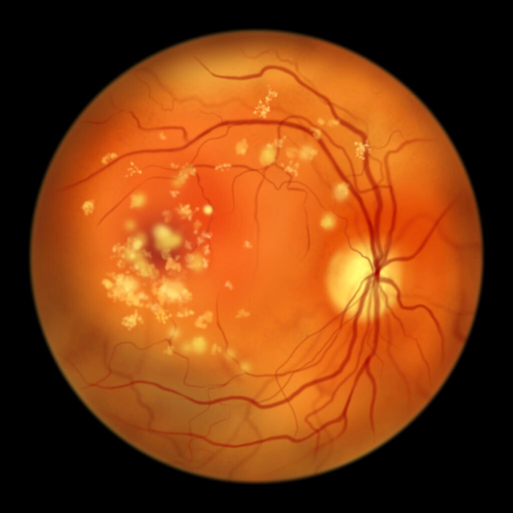
Drusen are small, yellowish deposits that collect between the retinal pigment epithelium (RPE) and a thin layer called Bruch’s membrane at the back of the eye. They're primarily made of lipids and proteins.
Drusen themselves typically aren't harmful. However, they often indicate age-related changes, such as age-related macular degeneration (AMD).
Drusen are routinely identified during AMD tests because certain drusen types signal a higher likelihood of progressing to advanced AMD. Fortunately, the presence of drusen alone doesn't mean immediate vision loss.
What Causes Drusen?
Drusen occur naturally with age. They result from the accumulation of proteins, lipids, and other unwanted material in the retina.
Usually, retinal pigment epithelial (RPE) cells recycle waste material. If their clearance pathways become overwhelmed or dysregulated, debris can accumulate as yellow deposits under the retina.
Who Is at Risk for Drusen?
Retinal drusen are commonly found in people over age 50. Caucasian people are more likely to develop drusen and age-related macular degeneration.
Larger drusen are a sign of AMD. Risk factors for AMD include:
- Family history of AMD
- High blood pressure
- High blood cholesterol levels
- Smoking tobacco products
What are the Different Types of Retinal Drusen?
Drusen are categorized mainly by size: small (<63 μm), intermediate (63–125 μm), and large (>125 μm). 'Hard' and 'soft' describe appearance; hard drusen are small with well-defined edges, while soft (often large) drusen are larger with indistinct edges.
The size of the drusen significantly impacts the risk of AMD development. Additionally, hard drusen have clear edges, whereas soft drusen appear fuzzy and indistinct, often merging together.
Soft drusen are larger than hard drusen and tend to cluster together. Having a lot of intermediate or large drusen is a major risk factor for vision loss.
What is Reticular Pseudodrusen?
Reticular pseudodrusen (subretinal drusenoid deposits) form above the RPE layer, unlike typical drusen. Their presence is strongly linked to an increased risk of progressing to late-stage AMD and associated vision loss.
Symptoms And When To Call Your Doctor
Most people with drusen experience no noticeable symptoms. Unlike AMD itself, drusen typically don't directly cause visual problems.
However, they warrant regular check-ups to detect early signs of potential AMD progression. Contact your eye doctor immediately if you experience:
- Vision distortion (straight lines appearing wavy or bent)
- Dark or gray spots in your central vision
- Blurry or hazy vision that worsens within days
- New floaters or flashes
- Difficulty seeing when transitioning from bright light to low light
How is Drusen Diagnosed?
Eye doctors detect drusen using a dilated eye exam. Dilating eye drops widen the pupils, allowing a thorough inspection of the retina.
Doctors often take baseline retinal photographs to monitor changes in drusen size and number during future visits. Additionally, advanced imaging technologies help track drusen more precisely:
- Optical coherence tomography (OCT). Provides cross-sectional retina views, clearly showing drusen size, shape, and retinal stress signs.
- Fundus autofluorescence (FAF). Highlights the health of RPE cells, indicating areas under stress or damage.
Lastly, you can use an Amsler grid at home to help detect early AMD-related changes. If you notice distorted or missing grid lines, or other changes to your vision, contact your eye care provider promptly.
How is Drusen Treated?
While drusen can’t be directly treated, there are ways to manage their impact and slow their progression. For instance, people with AMD may benefit from the AREDS2 vitamin and mineral supplement.
AREDS2 includes vitamins C and E, lutein, zeaxanthin, zinc, and copper, and has shown about a 25% reduction in AMD progression risk over five years.
Smokers should specifically choose the beta-carotene-free AREDS2 to avoid additional lung cancer risks.
Lifestyle and Dietary Changes
Consider switching to a healthier diet consisting of leafy greens, colorful vegetables, fruits, fish, nuts, and olive oil. This helps support retinal health and improve your overall health.
Quitting smoking also significantly reduces AMD risk. Additionally, regular physical activity alongside blood pressure control further aids retinal health.