

Heterochromia is when a person's eyes are not the same color, or when a single iris has more than one color. It is usually harmless and present from birth, but in rare cases it can be linked to an eye condition, an injury, or a genetic syndrome. There are three main types: complete heterochromia (each eye a different color), central heterochromia (a ring of a different color around the pupil), and segmental heterochromia (a patch or wedge of a different color within one iris). This article covers all three, plus when eye-color differences need to be checked by an eye doctor.
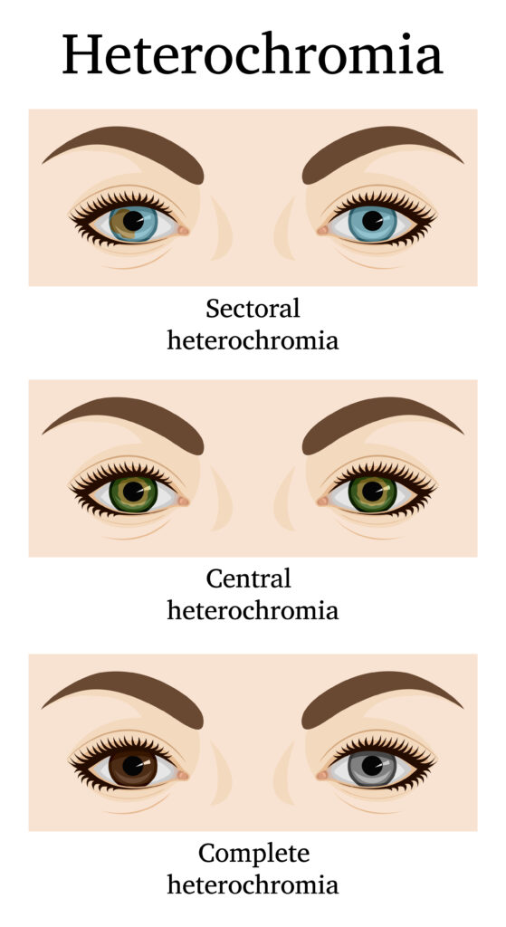
The Three Types of Heterochromia
Heterochromia is grouped by where the color difference appears. All three types are usually present from birth, and most are harmless.
| Type | What you see | How common |
|---|---|---|
| Complete heterochromia (heterochromia iridum) | Each eye is a clearly different color (for example, one blue eye and one brown eye). | Rare. One photo-based study estimated it at roughly 0.06 percent of people. |
| Central heterochromia (heterochromia iridis) | A ring of a different color around the pupil, surrounded by the main iris color. Usually appears in both eyes. | The most common form. Frequently mistaken for hazel eyes. |
| Segmental heterochromia (sectoral heterochromia) | A patch, wedge, or section of one iris is a different color from the rest. | Uncommon. Subtle and easy to miss unless you look closely. |
If your eyes have always looked this way, all three types are usually a benign genetic variation. If your eye color changed suddenly or only one eye is affected, see an eye doctor; color change in adulthood can signal an underlying condition.
Common Questions About Heterochromia
These short answers cover the questions readers search for most. The longer explanations follow below.
Does central heterochromia affect both eyes?
Central heterochromia usually appears in both eyes with the same color pattern in each iris. If the ring shows in only one eye, or the color appeared recently, get it checked. One-sided change points to a different cause.
What colors does central heterochromia usually show?
The most common pattern is a gold, amber, or hazel ring inside a blue, green, or grey outer iris. Brown irises also show a darker central ring, though the contrast is harder to see.
Is heterochromia always blue and brown?
No. Heterochromia appears in any combination of eye colors. Blue and brown is the most visually striking pairing and the one shown most in photos, but green-and-brown, hazel-and-blue, and other combinations all occur.
What is the difference between heterochromia iridum and heterochromia iridis?
In clinical writing, "heterochromia iridum" refers to color differences between the two eyes (complete heterochromia), and "heterochromia iridis" refers to color differences within a single iris (central or segmental). The two terms are used loosely and even interchangeably in medical literature.
How rare is heterochromia?
Exact numbers are not well established. One peer-reviewed study of high-resolution yearbook portraits estimated complete heterochromia at roughly 0.06 percent of people. Cleveland Clinic and StatPearls both describe heterochromia as rare but say the precise prevalence is unknown.
How do I know which type I have?
The Three Types table above is the fastest way to tell. If a ring of a different color surrounds your pupil in each eye, that is central heterochromia. If your two eyes are clearly different colors, that is complete heterochromia. If a wedge or patch of one iris is a different shade, that is segmental.
What is Heterochromia?
Heterochromia is the medical term for differences in eye color. It appears in three main forms, described below.
Central Heterochromia
Central heterochromia happens when the inner ring of the iris (around the pupil) is a different color from the outer part. The effect is a striking halo or two-toned look in each eye. A typical example is a gold or hazel ring near the pupil, surrounded by a blue or green outer iris.
This pattern usually appears in both eyes with the same color combination. It is a benign genetic variation in most people, not a sign of disease. Eye doctors describe it as a cosmetic difference that does not harm vision or eye health.
If you suddenly notice this color pattern appearing, or if it only shows in one eye, have it checked; several eye conditions mimic central heterochromia.
Complete Heterochromia
Complete heterochromia means each eye is a different solid color. For example, one eye is green and the other blue. This type is usually present at birth and does not affect vision or health.
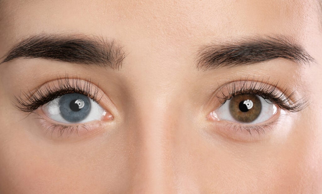
Woman with different colors of eyes, closeup. Heterochromia iridum.
In rare cases, complete heterochromia signals an underlying medical condition, especially when it appears suddenly later in life.
Segmental Heterochromia
Segmental heterochromia, also called sectoral heterochromia, means part of one iris has a contrasting color. You will see a slice, patch, or speckle of brown in a blue iris or vice versa.
This pattern is usually subtle and easy to miss unless you look closely. It is either a natural variation or the result of an eye condition or injury.
How Common Is Heterochromia?
Heterochromia is rare, but exact prevalence is not well established. One peer-reviewed photo-based study estimated complete heterochromia at roughly 0.06 percent of people. StatPearls notes there is no widely reported prevalence number for heterochromia overall, and Cleveland Clinic describes the condition as uncommon without a fixed figure.
Heterochromia is more visible in people with lighter base eye colors because contrast against blue, green, or grey shows more clearly than against brown.
What Determines Eye Color?
Your eye color is shaped by genetics, pigment, and how light scatters inside your eyes. Most of it comes down to melanin, the pigment that gives the iris its color.
People with more melanin have brown eyes. People with less melanin have blue, green, or hazel eyes. Hazel eyes shift in apparent color because they combine several pigment layers and scattering effects.
The type of melanin matters too. Brown-black eumelanin and red-yellow pheomelanin combine in varying amounts to produce different shades.
Blue eyes do not contain blue pigment. Light scatters through a low-pigment iris, making the eye look blue (the same scattering effect that makes the sky look blue).
Genes That Affect Eye Color
Dozens of genes shape eye color, including key genes on chromosome 15. These genes control how much melanin your eyes make and where it is stored.
That is why eye color varies so much, even among family members, and why some people have more than one color in a single iris.
Causes of Heterochromia
Heterochromia falls into one of three categories: congenital (present from birth, usually harmless), syndromic (linked to a genetic condition like Waardenburg or Horner syndrome), or acquired (developed later from injury, inflammation, glaucoma medication, or rarely a tumor). Most cases are the first kind.
Congenital Causes
Most heterochromia is congenital; you are born with it. It is a harmless variation in how melanin is distributed in the irises. It does not affect vision and does not need treatment.
Underlying Conditions
In some cases, heterochromia is linked to a genetic or medical condition, such as:
Waardenburg syndrome. A genetic condition that can cause different-colored eyes, white patches of hair, and congenital hearing loss in some people, though most people with the syndrome have normal hearing.
Sturge-Weber syndrome. Linked to a darker iris on the same side as a facial port-wine birthmark.
Horner syndrome. When present from birth, it produces a lighter-colored iris on the affected side.
Parry-Romberg syndrome. Causes one side of the face to change over time, including iris color.
Acquired Causes
Eye-color differences that develop later in life come from:
Eye injury. Trauma damages iris pigment or deposits iron in the eye (siderosis), altering its color.
Chronic inflammation. Long-term conditions like Fuchs uveitis syndrome (also called Fuchs heterochromic iridocyclitis) cause one iris to lighten gradually over years.
Glaucoma treatments. Prostaglandin-analog eye drops darken the iris over time as a known side effect.
Iris tumors. A new dark spot or freckle on the iris that grows, changes shape, or distorts the pupil is a sign of an iris nevus or, rarely, iris melanoma. Any new pigmented iris lesion should be checked.
Pigment dispersion. Pigment rubs off the back of the iris and settles on structures in the front of the eye. This shows up as see-through gaps in the iris, and the loose pigment raises eye pressure in some people; a condition called pigmentary glaucoma.
New or changing eye color should always be evaluated, even though most heterochromia is benign.
How Heterochromia Is Diagnosed
Most heterochromia is identified during a routine eye exam by comparing iris pigmentation under a slit lamp. If your eye color has changed recently or only one eye is affected, your doctor will also check for inflammation, eye pressure, and signs of injury.
Your eye doctor (usually an optometrist or ophthalmologist) will look closely at your irises, pupils, and overall eye health. During the exam, they will:
Check visual acuity to confirm your eyesight is normal
Compare iris pigmentation in both eyes, looking for unusual patterns or changes
Measure eye pressure to rule out glaucoma or other pressure-related conditions
Look for signs of inflammation or injury that could affect iris color
If your doctor suspects a medical condition, they will recommend imaging (such as OCT or ultrasound) to examine deeper eye structures, or blood tests to assess inflammation or genetic markers.
In children with other signs (like a drooping eyelid or unequal pupils), doctors often order scans or lab tests to rule out rare conditions such as neuroblastoma.
Even when everything looks healthy, your doctor will sometimes take a photo for future comparison.
When to See an Eye Doctor
Most heterochromia is harmless and does not require medical attention. Get same-day eye care if a new eye-color change comes with any of these symptoms:
Eye pain
Significant redness
Light sensitivity
New or sudden blurry vision, or any other vision change
A drooping eyelid or unequal pupil size, which can point to a nerve issue
Schedule a routine eye exam soon if you notice a new eye-color change without those symptoms, especially in just one eye.
In children, heterochromia combined with a drooping eyelid or unequal pupils warrants a pediatric ophthalmology referral to rule out rare conditions like neuroblastoma.
If your eyes have always looked this way and you have not noticed any recent changes, there is usually no cause for concern.
Treatment and Cosmetic Options
Heterochromia itself does not need medical treatment. The focus is on managing any underlying condition, if one is identified.
If your eye color change is due to chronic inflammation, your doctor will prescribe anti-inflammatory medication. If a tumor or pressure-related issue is involved, treatment targets that specific cause to protect your vision.
If your eyes are healthy and you simply prefer them to match, cosmetic options are available. The safest and most common choice is colored contact lenses. An eye care provider will prescribe:
Matching lenses to make both eyes look the same
Enhancement tints to highlight or balance color differences
Custom lenses for more dramatic or specific results
Makeup helps too. Some people use eyeshadow or eyeliner shades that accentuate their eye color or make it less noticeable.
Avoid cosmetic eye-color-change surgery. That includes iris implants, laser depigmentation, and keratopigmentation (also called corneal tattooing). None of these are FDA-approved for cosmetic eye-color change in the United States, and they have been linked to vision loss, glaucoma, chronic inflammation, corneal damage, and serious eye infections.
The American Academy of Ophthalmology recommends against cosmetic eye-color-change procedures. The only artificial iris device the FDA has approved is for medical iris defects (like a missing iris from a birth condition or injury), not cosmetic color change.
Some clinics outside the United States (notably in Panama, Turkey, and Iran) market iris implants directly to consumers despite documented vision-loss complications. Always talk to an eye doctor before trying to change your eye color.
Living With Heterochromia
If your eye color has always looked different and your vision is healthy, there is no medical reason for concern. Heterochromia is a cosmetic trait in most people, not a medical problem.
People with benign heterochromia have full vision and a normal life. Still, protect your eyes by:
Wearing sunglasses to reduce UV exposure
Using protective eyewear during sports and yard work to avoid injury
Getting regular eye exams to track any changes over time
Some people feel self-conscious about having different-colored eyes, especially when others comment on them. Many find it empowering once they understand the condition.
If you have questions about your eye color, or notice any new changes, the best next step is a comprehensive eye exam.
Summary
- Heterochromia is when the eyes are different colors, or when one iris has more than one color. The three types are complete, central, and segmental.
- Most cases are harmless and present from birth. No treatment is needed unless an underlying condition is causing it.
- Get same-day eye care if a new eye-color change comes with eye pain, significant redness, light sensitivity, or new vision problems. Otherwise, schedule a routine exam soon.
- Avoid iris implants, laser depigmentation, and keratopigmentation for cosmetic color change. None are FDA-approved for that purpose, and they have caused vision loss, glaucoma, inflammation, and corneal damage.

