

A bubble on the eye is a fluid-filled or tissue growth on the white of the eye (sclera) or the clear membrane covering it (conjunctiva). Six different conditions can cause it, ranging from harmless sun damage to a rare cancer. The bubble's color, location, and shape are the fastest way to narrow down which one you are looking at. Most bumps and bubbles develop on the conjunctiva, the thin outer layer of the eye.
| What you see | Likely cause | When to see a doctor |
|---|---|---|
| Pink or white wedge growing from the side of the eye toward the cornea | Pterygium ("surfer's eye") | Within a week if it is irritating; sooner if it is growing onto the cornea |
| Yellowish patch near the nose-side of the eye | Pinguecula | Routine eye exam |
| Clear, fluid-filled blister on the white of the eye | Conjunctival cyst | Within a week; sooner if painful |
| Solid lump where the cornea meets the white of the eye, present since birth | Limbal dermoid (congenital, noncancerous) | Pediatric ophthalmology referral |
| Reddish, white, brown, salmon-colored, or rapidly growing spot | Conjunctival tumor (may be cancerous) | Same week; biopsy may be needed for suspicious lesions |
| Sudden swelling of the clear membrane that looks like a blister or bag of fluid | Chemosis (usually from allergy, infection, or trauma) | Same day if vision is affected; call 911 or go to an ER right away if swelling comes with trouble breathing, fainting, or swelling of the lips, tongue, or face |
If your eye is also painful, you have vision changes, or the bubble appeared suddenly after an injury, see an eye doctor or visit an emergency room the same day. If you are looking for general eye anatomy rather than a growth or symptom, see Eye Anatomy: Parts of the Human Eye.
6 Causes of a Bubble on the Eye
1. Pterygium
A pterygium is a wedge-shaped tissue growth on the conjunctiva that starts on the side of the eye. It can keep growing toward the cornea, cause discomfort, and even affect vision.
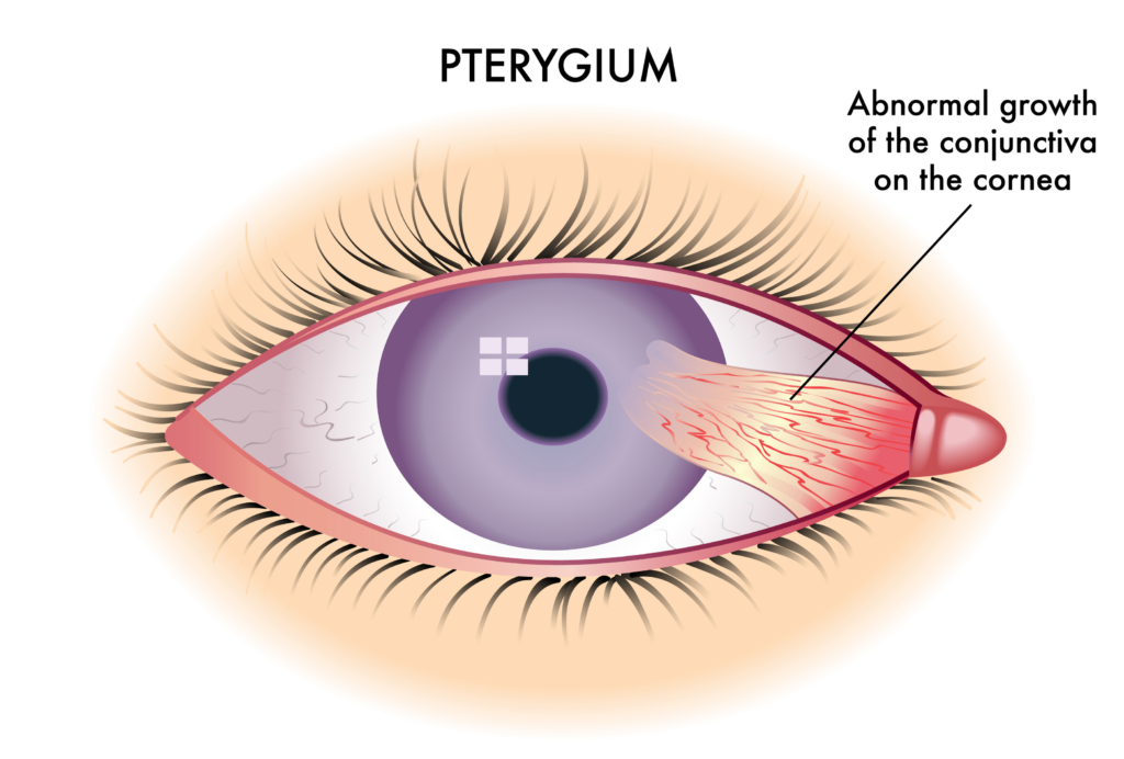
More common names for pterygium include "surfer's eye" or "farmer's eye." People who live in sunny, arid, or dusty environments are more prone to pterygium.
Symptoms of a pterygium include:
- Dry eye
- Blurred vision or astigmatism
- Pink or white wing-shaped growth on the eye
Listen In Q&A Format
What Causes Bubbles or Bumps on Your Eyeballs?
Vision Center Podcast
2. Pinguecula
A pinguecula is a yellowish patch or bump that usually appears on the side of the eye nearest the nose. It is a deposit of protein, fat, or calcium that develops from chronic irritation.
Common causes of pinguecula include:
- The aging process
- Exposure to UV light
- Dry eyes
Symptoms of pinguecula include:
- Burning or stinging eyes
- Dry or red eyes
- Itching eyes
- Tearing
- Inflammation or swelling
- Foreign body sensation (feeling like something is in your eye)
- Blurred vision
A pinguecula that grows and extends across the cornea becomes a pterygium. At that stage it can affect vision, and your eye doctor may recommend surgical removal if it changes the shape of the cornea, causes ongoing irritation, or threatens sight.
3. Conjunctival Cyst (Clear Bubble)
A conjunctival cyst is a thin-walled, benign sac that holds clear fluid. It sits on the conjunctiva, the membrane that covers the white of the eye and lines the inside of the eyelid. Most are harmless and often resolve on their own.
A conjunctival cyst can be congenital (present from birth) or develop later after inflammation, trauma, or eye surgery. If a cyst causes symptoms, an eye doctor can drain or remove it in-office.
If you have a conjunctival cyst, you may notice:
- Eye redness or irritation
- Increased tear production
- Discomfort or a foreign body sensation
4. Limbal Dermoid (Congenital Growth)
A limbal dermoid is a benign congenital growth (present from birth) found where the cornea and sclera meet. It appears as a solid yellow-white mass, not a cyst. The medical term is epibulbar choristoma (normal tissue growing in an abnormal location).
A limbal dermoid can grow large enough to affect vision. It does not have a single specific cause, but it is often associated with other ocular and systemic conditions, including:
- Goldenhar syndrome
- Duane's syndrome
- Coloboma of the upper lid
- Lacrimal stenosis
Small, symptom-free limbal dermoids are often watched over time with pediatric ophthalmology follow-up. Surgery comes into play when the growth threatens vision, causes ongoing irritation, interferes with eyelid closure, or is large enough to be a significant cosmetic concern.
5. Conjunctival Tumor
Some conjunctival tumors are cancerous and need a biopsy to confirm. The most common cancerous forms are squamous cell carcinoma, conjunctival melanoma, and lymphoma, all of which require an eye doctor's evaluation.
Squamous Cell Carcinoma
This tumor appears reddish or white, and flat or elevated. Squamous cell carcinoma of the conjunctiva rarely spreads to distant parts of the body, but it can extend into the eye orbit and sinuses and damage vision.
Conjunctival Melanoma
Conjunctival melanoma can develop from primary acquired melanosis, from a pre-existing nevus (eye freckle), or on its own. Any change in size, color, or shape of a pigmented spot on the eye should be checked by an eye doctor. Treatment usually involves surgical removal.
Lymphoma
This salmon-colored lesion often hides on the eye surface beneath the eyelid. It may be limited to the conjunctiva or be a sign of systemic lymphoma. Your eye doctor will arrange the work-up needed to confirm, which may include imaging or a biopsy.
6. Chemosis
Chemosis is sudden swelling of the conjunctiva that looks like a blister or a bag of fluid on the white of the eye. It happens when fluid builds up under the membrane, usually as a reaction to allergy, infection, or trauma.
Common triggers include:
- Allergies
- Bacterial or viral infections
- Hyperthyroidism (thyroid eye disease)
- Eye trauma or rubbing
- Surgical complications
Symptoms of chemosis include:
- Itchy eyes
- Eye irritation
- Excessive tearing
- Puffy eyes
- A water-balloon-like swelling on the white of the eye
Chemosis with eye pain or vision change needs same-day medical care. If the swelling comes with trouble breathing, fainting, or swelling of the lips, tongue, or face, call 911 or go to an emergency room right away.
Diagnosis
Diagnosis usually starts with a visual exam by an optometrist or ophthalmologist. Many bubbles on the eye can be identified by appearance alone. For pigmented, rapidly changing, or atypical growths, your eye doctor may use photo-documentation, imaging, or a biopsy to track or confirm the diagnosis.
During the exam, your eye doctor will ask about:
- Eye injuries or problems you have had
- Your contact lens wearing habits, if you wear contacts
- Cosmetics, eyelash extensions, and other products that could irritate your eyes
Treatment by Condition
Treatment depends on the cause. Pterygium and pinguecula often need only artificial tears and UV protection. Cysts and limbal dermoids may need monitoring or surgical removal. Conjunctival tumors require evaluation and may need surgery, radiation, or chemotherapy.
- Pterygium and pinguecula: Artificial tears, UV-blocking sunglasses, and short courses of steroid or anti-inflammatory eye drops for irritation. Surgery is an option when the growth causes ongoing irritation, affects vision, changes the shape of the cornea, or creates a significant cosmetic concern. Pterygium can recur after surgery, and recurrence risk varies by surgical technique.
- Conjunctival cyst: Most resolve on their own. Persistent cysts can be drained or surgically removed in-office, depending on size and symptoms.
- Limbal dermoid: Treatment depends on size and how the growth affects vision. Small, symptom-free dermoids are often monitored with pediatric ophthalmology follow-up. Surgery is used when the dermoid threatens vision, causes significant irritation, affects eyelid closure, or is a major cosmetic concern.
- Conjunctival tumor (cancerous): Treatment depends on biopsy results and may include surgical excision, cryotherapy, topical chemotherapy eye drops (mitomycin C, interferon, or 5-fluorouracil), radiation, or immunotherapy. Ocular oncology referral is the standard pathway.
- Chemosis: Treat the underlying cause: antihistamines for allergy, antibiotics for bacterial infection, removal of the irritant for trauma. Cool compresses help reduce swelling.
Home Care for Pterygium and Pinguecula
You can reduce irritation and slow growth with a few simple habits:
- Wear UV-blocking wraparound sunglasses whenever you are outside; UV-blocking contact lenses can add protection but do not replace sunglasses
- Use wraparound glasses, goggles, or other protective eyewear in dry, dusty conditions
- Use artificial tears frequently to prevent dryness in arid environments
Risks of Eye Bubbles
Most bubbles on the eye are benign, but some carry real risks if ignored:
- Vision impairment, such as astigmatism from a large limbal dermoid or a pterygium that reaches the cornea
- Cancerous conjunctival tumors can invade nearby tissues or spread, so suspicious growths need timely evaluation and ocular oncology referral
- A pterygium can develop from an untreated pinguecula in patients with continued sun and dust exposure
- Secondary infection of an irritated or open lesion
- Amblyopia (lazy eye) in children with a vision-blocking limbal dermoid
- Eye scarring after surgery or chronic inflammation
Early evaluation by an eye doctor is the best way to prevent vision loss and complications.
When to See an Eye Doctor
Most bubbles on the eye are not emergencies, but some need same-day attention. See an eye doctor or visit an emergency room the same day if you have any of the following:
- The bubble appeared suddenly, especially after an injury, a sneeze, or coughing
- The bubble is growing, changing color, or has a brown, dark, or salmon-colored spot
- You have eye pain, light sensitivity, or vision changes
- The bubble is on a child's eye and was not there before, or has been present since birth (limbal dermoid needs pediatric ophthalmology referral)
- The white of the eye looks swollen like a water balloon. If this happens with trouble breathing, fainting, or swelling of the lips, tongue, or face, call 911 or go to the emergency room.
For non-urgent cases, such as a stable yellowish patch (pinguecula) or a wedge growth that is not changing, see your eye doctor at your next routine exam.
Summary
A bubble or blister on the eye can be a pterygium, pinguecula, conjunctival cyst, limbal dermoid, conjunctival tumor, or chemosis. The color, location, and onset of the growth help narrow the diagnosis. Most are benign and manageable, but any rapidly growing, painful, or suspicious spot needs prompt evaluation by an eye doctor.

