If your eye doctor has mentioned phototherapeutic keratectomy (often called PTK), you may be wondering what exactly it involves.
Laser surgery on your eyes sounds serious, especially when words like “scarring” or “corneal erosion” are used in the same sentence. The good news is that PTK is a medically guided, low-risk procedure designed to treat specific problems on the surface of your eye.
In this guide, we’ll discuss what PTK is, what it treats, how it’s different from other laser surgeries, and what to expect before, during, and after the procedure.
What is Phototherapeutic Keratectomy?
Phototherapeutic keratectomy (PTK) is a laser treatment that removes damaged or diseased tissue from the front layers of your cornea, the clear surface of your eye. By carefully smoothing this area, PTK can relieve symptoms such as pain, blurred vision, and recurring surface breakdowns.
PTK uses a medical laser called an excimer laser, which emits ultraviolet light at a precise wavelength. This laser gently vaporizes tissue in micro-thin layers. Because of its precision, PTK is ideal for treating superficial problems without damaging deeper structures.
Doctors often describe PTK as a bridge between medical treatments (like eye drops) and major surgeries (like a corneal transplant). It doesn’t work for every eye condition, but when it’s the right fit, it can make a big difference with fewer risks than more invasive procedures.
Conditions That PTK Can Treat
PTK is designed to treat specific surface-level eye conditions. It’s most effective when the problem affects the outermost layer of the cornea and hasn’t damaged deeper tissue.
Here are the types of conditions PTK is often used to treat:
Corneal dystrophies. Inherited conditions like granular, lattice, or Reis-Bücklers dystrophy that cloud the cornea with deposits.
Recurrent corneal erosions. A condition where the surface of the eye repeatedly breaks down, causing sharp pain and light sensitivity.
Superficial corneal scars. These may result from infections, injury, or past surgeries that left cloudy patches on the surface.
Band keratopathy. Calcium buildup across the front of the eye that creates a white, gritty band and blurs vision.
Salzmann’s nodular degeneration. Blue-white nodules on the cornea that distort shape and vision.
Bullous keratopathy (for symptom relief). When the inner corneal layer fails, PTK can help reduce surface pain even if it doesn’t restore vision.
PTK does not treat deep corneal scars or endothelial diseases like Fuchs’ dystrophy. It isn’t used for active infections either.
If your eye doctor has diagnosed one of the conditions above, it’s best to ask whether PTK might be appropriate for your case.
PTK vs. Other Laser Surgeries
PTK may use the same laser technology as other eye surgeries, but it serves a different purpose. Understanding how it compares helps set the right expectations.
Here’s how PTK stacks up against similar procedures:
- PTK. Removes disease or scars from the corneal surface to improve comfort and clarity of vision—not primarily to eliminate the need for glasses.
- PRK (Photorefractive Keratectomy). Reshapes the cornea to correct vision problems like nearsightedness or astigmatism.
- LASIK. Involves creating a flap in the cornea before reshaping it with a laser; this enables rapid recovery but introduces flap-related risks.
- Superficial Keratectomy (SK). A manual scraping of surface lesions—sometimes done before or instead of PTK.
- Corneal Transplants. Replace deeper or fully damaged corneas; used when PTK is insufficient.
PTK is flapless, which means fewer complications related to corneal healing. It also may remove less tissue than PRK, depending on how much tissue needs to be removed, which can be helpful when corneal thickness is limited.
While it can’t replace a transplant for deep disease, it may delay the need for one.
Who is a Good Candidate for PTK?
PTK is most effective for surface-level problems in otherwise healthy eyes. Ideal candidates have:
Corneal opacities or deposits limited to the front 10–20% of the cornea.
Good overall eye health and stable vision needs.
No significant thinning or bulging of the cornea (ectasia).
The procedure is often recommended for people with recurrent erosions, superficial scars, or early-stage dystrophies that haven’t responded to drops or ointments.
How Doctors Decide on PTK
Choosing PTK is never based on a single exam—it requires a comprehensive evaluation to ensure the treatment is safe and likely to help.
The process usually begins with a slit-lamp exam, where your eye doctor uses a microscope and bright light to inspect your cornea. This helps them identify surface issues such as scars, dystrophic deposits, or erosions.
Next, several imaging tests help guide the plan:
Corneal topography maps the surface shape of your cornea to identify irregular curves or distortions.
Pachymetry measures corneal thickness to ensure sufficient tissue remains after treatment.
Optical Coherence Tomography (OCT) provides cross-sectional images that show the extent of the problem.
If the issue reaches too far into the cornea—or if your cornea is already thin—your doctor may recommend a different surgery.
Doctors will also screen for risk factors that could delay healing or increase the risk of complications. These include:
Uncontrolled diabetes or autoimmune diseases
History of herpes simplex virus in the eye
Severe dry eye or inflammation
Signs of corneal ectasia (a weak, bulging cornea)
In most cases, PTK is offered only after the eye is free of infection and stable enough to heal well after the procedure.
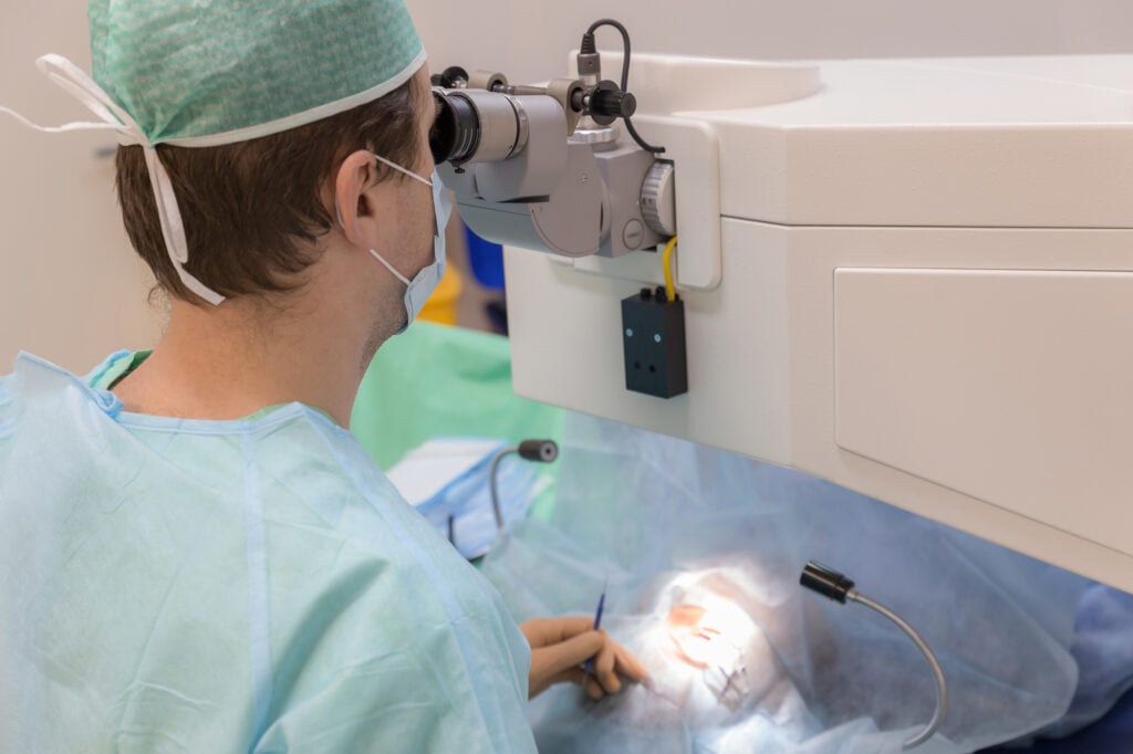
What is the PTK Procedure Like?
The PTK procedure is short and outpatient—meaning you’ll go home the same day. But knowing what to expect can make the day less stressful.
Here’s what usually happens:
Before surgery. You’ll stop using eye makeup or lotions a few days in advance. Your doctor may prescribe antibiotic drops and ask about any medications, especially blood thinners. Arrange a ride, as you won’t be able to drive afterward.
During the procedure. You’ll sit in a reclining chair while anesthetic drops numb your eye. A device gently holds your eyelids open. The surgeon removes the surface layer (epithelium) using a laser or a fine instrument.
Laser treatment. The excimer laser delivers pulses that remove only diseased tissue. If the surface is rough, a clear gel may be used to highlight high spots and guide smoother ablation.
After the laser. A bandage contact lens is placed on your eye to protect it while the surface heals. You’ll also start using prescribed antibiotic and anti-inflammatory drops.
The procedure typically takes 10 to 20 minutes per eye. You may notice some haziness or mild discomfort right away, but pain and light sensitivity are common once the numbing drops wear off, especially during the first few days.
Recovery and Self-Care After a PTK Procedure
The first few days after PTK are often the most uncomfortable, but proper care can help ease symptoms and support smooth healing. You’ll likely have a follow-up visit within 24 hours, then again at one week and one month.
During early recovery, you may notice hazy vision, light sensitivity, or a gritty feeling in your eye. These symptoms usually improve within a few days as your corneal surface heals.
First Days
The corneal surface begins to regenerate within 3 to 7 days. During this time, you’ll wear a soft bandage contact lens to protect your eye and reduce discomfort. Your doctor will prescribe:
Antibiotic drops. To prevent infection while the surface is still healing.
Pain relief. Often includes a cold compress, oral medications, or short-term anti-inflammatory eye drops.
Artificial tears. To keep your eyes lubricated and reduce stinging.
You’ll also be advised to rest, limit screen time, and avoid rubbing your eyes.
First Months
Even after the bandage lens is removed, your vision may continue to shift for a few weeks as your cornea remodels. It’s normal for your eyesight to fluctuate during this phase, especially in the first month.
Some people experience temporary glare, dry eye, or halos around lights. Artificial tears and avoiding wind or dry environments can help reduce these symptoms.
If you wore glasses before PTK, you may need a new prescription once your vision stabilizes.
Here are a few key things to avoid during recovery:
Rubbing your eyes. This can interfere with healing or dislodge the bandage lens.
Swimming or hot tubs. To prevent infection while the surface is still fragile.
Contact sports. Protect your eyes from accidental trauma.
Makeup or face creams near your eyes. Wait for your doctor’s approval before resuming.
Heavy lifting or dusty environments. These can increase irritation or risk.
Most people can resume routine activities like reading or desk work within a few days. For higher-risk activities, follow your doctor’s personalized guidance.
If your symptoms worsen instead of improving—or if you notice sudden pain, redness, or decreased vision—call your eye doctor right away.
When to Seek Help After PTK
You never have to “wait and see” alone after PTK. If something feels off, reaching out early is always the safer choice.
Watch for red flags such as:
Sudden or severe eye pain
Rapid decline in vision
Redness that worsens instead of improves
Discharge or crusting around the treated eye
These may signal infection or other complications and need same-day care.
Otherwise, follow your doctor’s timeline for follow-up visits—typically at 1 day, 1 week, and 1 month. If your condition recurs, another PTK may be an option, or you may be advised to consider a different treatment.
Store your clinic’s contact info in your phone and know how to reach them after hours. Most PTK recoveries go smoothly, and staying in touch with your eye care team helps keep it that way.
Risks, Side Effects, and Limits of PTK
Like any medical procedure, a PTK procedure carries risks. Most side effects are temporary, but it’s important to understand what to expect and when to seek help.
Most people experience some of the following during the first days or weeks:
Eye pain or discomfort. Especially during the first 48 hours.
Light sensitivity and glare. Often improves as healing continues.
Dryness or tearing. Can last for a few weeks to a few months.
Hazy vision or halos. Usually fades as the corneal surface smooths.
These effects are usually manageable with lubricants and medications. Follow-up visits help track healing and manage symptoms.
Serious Risks
Although uncommon, PTK can lead to:
Infection. Especially if the bandage lens is contaminated or healing is delayed.
Corneal haze or scarring. More likely with deeper treatments.
Reactivation of herpes simplex virus. If you’ve had eye herpes in the past.
Excessive thinning or corneal instability (ectasia). Rare, but possible if too much tissue is removed.
Delayed healing. Especially in people with diabetes, dry eye, or prior surgeries.
In people with inherited dystrophies, the same corneal changes may return months or years later. In many cases, repeated PTK can provide similar benefits, though repeat treatments may increase risks such as haze or refractive changes.
If you notice severe pain, sudden vision changes, or heavy discharge, contact your surgeon right away—it could signal an infection or other urgent issue.
PTK Costs, Insurance, and Access
Because PTK is performed for medical reasons—not for cosmetic vision correction—it’s often covered by insurance, but coverage varies by plan and indication. That may include Medicare and many commercial plans, provided the diagnosis and need meet policy criteria.
Conditions that often qualify include:
Corneal dystrophies and degenerations
Recurrent corneal erosion syndrome
Superficial scarring or irregularities
Band-shaped keratopathy (calcium deposits)
Most insurance plans require that conservative treatments (like eye drops or debridement) have already been tried without lasting success. You may also need to show that the problem is affecting vision or daily function.
If you don’t have insurance or the case isn’t covered, PTK can cost a few thousand dollars per eye. This is similar to PRK or LASIK pricing, since it uses the same laser systems.
Ask your eye surgeon’s office for billing codes and for help contacting your insurance provider in advance.